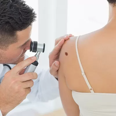
Dermoscopy Mole Evaluation - Abu Dhabi - Dubai
Dermoscopy is a non-invasive technique used to examine skin lesions, particularly moles, in greater detail. It involves using a dermatoscope, which is a handheld device equipped with a magnifying lens and a light source, to visualize the skin lesion's structures and patterns that may not be visible to the naked eye.
Dermoscopy helps dermatologists and other healthcare professionals evaluate moles and determine whether further investigation or intervention is necessary. Here are some key aspects that are assessed during dermoscopy mole evaluation:
- 1. ABCDE Criteria: Dermoscopy allows for the evaluation of specific features using the ABCDE criteria:
- • Asymmetry: Assessing the symmetry of the lesion. Asymmetrical moles may indicate potential malignancy.
- • Border irregularity: Examining the borders of the mole. Irregular or poorly defined borders can be a sign of malignancy.
- • Diameter: Measuring the size of the mole. Larger moles, typically greater than 6 millimeters, warrant closer examination.
- • Evolution: Monitoring changes in the mole over time. Any significant changes may indicate potential malignancy.
- 2. Structure and Patterns: Dermoscopy allows for the assessment of various structural elements and patterns within moles, including:
- • Pigment network: Examining the distribution and arrangement of pigmented lines or networks within the mole.
- • Dots and globules: Analyzing the presence of small dots or globules within the mole, which can indicate various characteristics.
- • Streaks: Observing linear or curved streaks within the mole, which may suggest atypical or abnormal growth.
- • Blue-white structures: Identifying areas with a blue-white appearance, which can be indicative of malignancy.
- • Vascular structures: Assessing the presence of blood vessels or other vascular patterns within the mole.
- 3. Additional Features: Dermoscopy can also reveal other features that may be relevant in evaluating moles, such as:
- • Regression structures: Identifying signs of regression, such as hypopigmentation or white scar-like areas.
- • Ulceration: Detecting areas of ulceration or erosion within the mole, which can raise concerns.
- • Surrounding skin: Assessing the skin surrounding the mole for signs of inflammation, redness, or other abnormalities.
It's important to note that dermoscopy is a tool used to assist in the evaluation of moles and skin lesions. A dermatologist or a healthcare professional with expertise in dermoscopy should interpret the findings and make a diagnosis. If any suspicious features are observed during dermoscopy, a biopsy may be recommended for further evaluation.