Gynecological ultrasound - Abu Dhabi - Dubai
Gynecological ultrasound, also known as pelvic ultrasound or pelvic sonography, is a medical imaging technique used to visualize and evaluate the female reproductive organs. It utilizes high-frequency sound waves to create real-time images of the pelvic region, including the uterus, ovaries, fallopian tubes, cervix, and surrounding structures. Here are some important points about gynecological ultrasound:
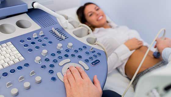
During a gynecological ultrasound, a transducer is used to emit sound waves into the pelvic region. The transducer can be placed on the abdomen (transabdominal ultrasound) or inserted into the vagina (transvaginal ultrasound) for better visualization of the pelvic organs.
Here are some common reasons why gynecological ultrasound may be performed:
- 1. Evaluating pelvic pain: Ultrasound can help identify the cause of pelvic pain, such as ovarian cysts, fibroids, or endometriosis.
- 2. Assessing abnormal bleeding: It can aid in diagnosing the cause of abnormal uterine bleeding, including conditions like uterine fibroids, polyps, or endometrial hyperplasia.
- 3. Monitoring pregnancy: Ultrasound is routinely used during pregnancy to assess fetal development, confirm gestational age, detect multiple pregnancies, and identify any potential abnormalities.
- 4. Diagnosing ovarian cysts: Ultrasound can provide detailed information about the size, location, and characteristics of ovarian cysts, helping in their diagnosis and management.
- 5. Detecting uterine abnormalities: Ultrasound can identify conditions like uterine fibroids, uterine septum, or congenital uterine anomalies, which may contribute to infertility or recurrent miscarriages.
- 6. Guiding procedures: Ultrasound can assist in guiding minimally invasive procedures such as biopsies, aspiration of cysts, or the placement of intrauterine devices (IUDs).
Gynecological ultrasound is generally safe, non-invasive, and does not involve exposure to ionizing radiation. The choice between transabdominal and transvaginal ultrasound depends on factors such as the patient's symptoms, the need for better visualization, and the stage of pregnancy if applicable. The procedure is typically performed by a trained sonographer or a gynecologist who interprets the ultrasound images to make a diagnosis or determine the next steps in treatment.
It's important to note that while ultrasound is a valuable tool, it has its limitations. In certain cases, additional imaging modalities or diagnostic tests may be required for a comprehensive evaluation of gynecological conditions. It is always recommended to consult with a healthcare professional for personalized advice and interpretation of ultrasound findings.